Gross image showing dilated and thick-walled blood vessels with
$ 7.99 · 4.9 (72) · In stock


Thick walled blood vessels. F.

Photomicrographs of the lesion. A -Hematoxylin & eosin (H&E) stain
Diagnostics, Free Full-Text

Approach to Cystic Lesions in the Abdomen and Pelvis, with

Capturing the often-elusive diagnosis of idiopathic myointimal

Ashley STUECK Hepatobiliary & Gastrointestinal Pathologist
Gross image showing dilated and thick-walled blood vessels with

Photomicrograph of a pathological specimen revealing dilated

Interspersed varying sized blood vessels ranging from small thin
Diagnostics, Free Full-Text

Structure and Function of Blood Vessels

Cystic duct, Radiology Reference Article

Photomicrograph showing a cavernous angioma containing abnormal
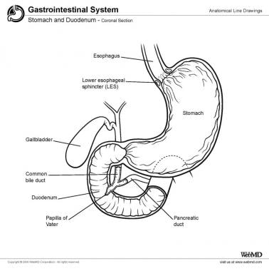
Duodenal Anatomy: Overview, Gross Anatomy, Microscopic Anatomy